The placement of a ureteric stent is not unusual in transplant surgery. Surgeons differ in their belief as to whether a stent offers any advantage. Some surgeons use a stent on all occasions, some never do, and some do selectively. Stents placed at the time of transplant are inserted by the surgeon such that they connect the pelvis of the kidney with the bladder by traversing the ureter. Stents should be removed 4-6 weeks following the surgery. At a minimum, the placement of the stent is recorded in the operative note dictated by the surgeon and in the nurses’ operative record. The patient must be informed of the stent placement, along with the risks and benefits of a stent, and provided information regarding the need and timing of its removal. In addition, the surgeon must inform the transplant team (e.g., nephrologists, transplant coordinators) that a stent has been placed so that it can be documented and a plan made for its removal. Complications from leaving a stent in place in a transplanted kidney include recurrent infections (bacterial, fungal), stone formation, mineralization, and obstruction of the kidney. Kidney infections in an immunocompromised patient can be life-threatening and result in significant damage to the kidney. Kidney infections resulting from prolonged stent placement cannot thus be eradicated by antibiotics alone. Because the stent is a foreign body, it will harbor the bacteria, preventing the antibiotics from effectively treating the infection.
Ureteric stent placement medical expert witness specialties include urology, pediatric urology, nephrology, pediatric nephrology, emergency medicine, infectious disease, pediatric infectious disease, and transplant urology.
IF YOU NEED AN Ureteric Stent Placement MEDICAL EXPERT, CALL MEDILEX AT (212) 234-1999.
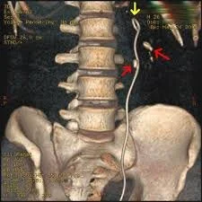
Image courtesy of Steven Fruitsmaak.Description Three-dimensional reconstructed CT scan image of a ureteral stent (left kidney, indicated by yellow arrow) in a 26-year-old male. There is a kidney stone in the pyelum of the lower pole (highest red arrow) and one in the ureter beside the stent (lower red arrow).
This file is licensed under the Creative Commons Attribution-Share Alike 3.0 Unported license.

